








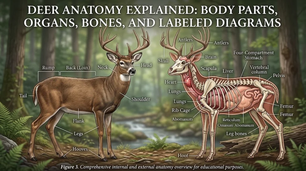
Deer anatomy is built for browsing, digesting tough plant material, sensing danger, and moving quietly through woodland, grassland, and mountain habitats. Their long legs, cloven hooves, flexible neck, large ears, strong senses, antlers, and ruminant digestive system all work together.
This guide explains the anatomy of a deer in simple language. It includes an external body-parts diagram, an internal-organ diagram, a short look at the skeleton, and the main differences readers notice between deer species.
External Deer Anatomy
Head, muzzle, eyes, and ears
A deer’s head is shaped for feeding and awareness. The muzzle helps the animal select leaves, shoots, grasses, fruit, and other plant foods. Deer do not have upper incisors like humans. Instead, the lower front teeth press against a tough dental pad in the upper jaw, helping the animal pull and cut vegetation. [1]
The eyes sit on the sides of the head, giving deer a wide field of view. That arrangement is useful for a prey animal that needs to notice movement from different directions. The ears can rotate and help the deer locate sounds, including sounds made by predators or people.
Neck, shoulder, and chest
The neck connects the head to the shoulders and allows the deer to reach vegetation at different heights. The shoulder and chest contain strong muscles that support the front of the body and help with running, jumping, and climbing uneven ground.
A deer’s body is not shaped like a horse’s body in every detail. Deer are even-toed hoofed mammals, and their limbs are adapted for a fast, springy stride. The exact proportions differ between roe deer, red deer, reindeer, moose, white-tailed deer, and other members of the deer family.
Legs, joints, and hooves
Deer legs look slim, but the lower limbs are specialized for movement. The forelegs support the front of the body, while the hind legs provide much of the pushing power during a run or jump. The joints and lower-leg bones align to create an efficient stride.
Each hoof is split into two main toes. Small dewclaws sit higher on the back of the foot and may leave marks in soft mud or snow. A track can therefore provide useful evidence about deer activity, but track shape alone should not be used to identify the exact species in every situation.
Rump and tail
The rump connects the spine to the hind limbs and contains large muscles used during acceleration. The tail differs noticeably between species. White-tailed deer are known for raising a bright white underside when alarmed, while mule deer have a smaller tail with a black tip. [2]
Antlers and the Deer Skull
Antlers are one of the most recognisable parts of deer anatomy. They are true bone that grows as an extension of the skull. In most deer species, males grow antlers, shed them, and regrow them in a yearly cycle. During growth, antlers are covered by a living skin called velvet. [3]
Antlers are not the same as horns. Horns have a permanent outer sheath and are generally not shed annually. Antlers are single bony structures and are normally shed after the growth cycle ends. [3]
Antler shape can help people discuss a species or age class, but it is not a complete identification method. Young deer, females of some species, and deer at different points in the annual cycle may not match a typical picture.
Deer Anatomy Diagram: Internal Organs
Heart and lungs
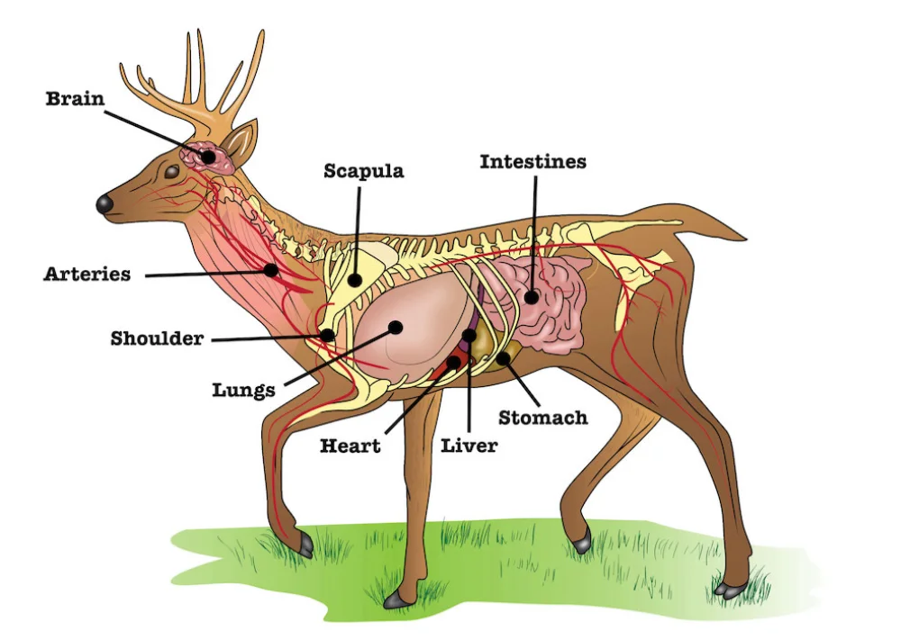
The heart pumps blood through the body. The lungs exchange oxygen and carbon dioxide as the deer breathes. Together, the heart, lungs, rib cage, and breathing muscles support activity such as walking, escaping danger, and seasonal movement.
The rib cage helps protect organs in the chest. It also provides attachment points for muscles involved in breathing and movement. A diagram of deer anatomy usually places the lungs behind the front ribs and the heart between them, slightly toward the front of the abdominal area.
Liver and intestines
The liver performs several essential functions, including processing nutrients and producing substances involved in digestion. The intestines absorb nutrients and water after food has passed through the stomach compartments.
The exact shape and position of organs varies with species, body size, age, and whether the digestive tract is full. A simplified deer anatomy chart is useful for orientation, but it should not be treated as an exact dissection map.
The four-compartment stomach
Deer are ruminants. Their stomach is commonly described through four compartments: the rumen, reticulum, omasum, and abomasum. [4]
- Rumen: the largest fermentation chamber, where microbes help break down plant material.
- Reticulum: works with the rumen and sorts smaller and larger particles.
- Omasum: helps absorb water and some nutrients while moving food onward.
- Abomasum: the glandular compartment that works most like the simple stomach people imagine.
After grazing or browsing, a deer can bring partly digested food back up and chew it again. This process is called rumination. It allows the animal to gain more nutrition from fibrous vegetation than a simple-stomach animal could obtain from the same food.
Deer Anatomy Bones and Skeleton
The deer skeleton includes a skull, vertebral column, ribs, pelvis, and long bones in the limbs. The spine supports the body and transfers movement from the hindquarters through the torso. Ribs protect the chest and help form the body cavity.
The pelvis connects the spine to the hind limbs. Strong muscles around the pelvis and thighs help the deer accelerate and jump. The lower limb bones are elongated and arranged for efficient movement rather than grasping.
White-tailed deer osteology and other deer skeleton studies show why a species-specific skeleton reference matters. A moose skeleton, a roe deer skeleton, and a white-tailed deer skeleton share the same basic mammalian plan but differ in size and proportions. [5]
Whitetail Deer Anatomy Compared With Other Deer
“Whitetail deer anatomy” usually refers to the white-tailed deer, a species in the genus Odocoileus. Its most familiar external features include a large tail with a white underside, long legs, a brown seasonal coat, and antlers in males.
Mule deer are related but have different identifying features, including larger ears and a black-tipped tail. Reindeer are also members of the deer family, but their antler pattern, body form, and adaptations reflect their colder habitats. The word “deer” describes a family of species, not one identical body plan.
When using a deer anatomy drawing or anatomy of a deer diagram online, always check which species it represents. A labeled illustration made for a white-tailed deer may not show every feature in a reindeer, roe deer, red deer, or moose.
Female Deer Anatomy and Fawns
A doe has the same basic skeletal, muscular, respiratory, and digestive systems as a buck. The visible differences people notice most often involve antlers and reproductive anatomy. In many species, adult males grow antlers while females do not, although there are exceptions.
Fawns have the same basic body plan as adults but smaller proportions and developing bones and muscles. White spots in the coat help camouflage many young deer. Because age and species change appearance, a single photograph should be interpreted carefully.
Why Deer Anatomy Matters in Wildlife Study
Learning anatomy helps readers understand behaviour. A deer’s hooves explain why it can cross rough ground. Its ears and side-positioned eyes explain how it reacts to sound and movement. Its ruminant stomach explains why it spends time feeding, resting, and chewing cud.
Anatomy also connects to ecology. Deer browse plants, move through habitats, and influence vegetation. Our guide to Norwegian animals and their role in nature explains how herbivores fit into wider ecosystems.
If you observe deer, keep a respectful distance, avoid feeding them, and do not chase them for a better photograph. See our guide to responsible wildlife watching in Norway for practical observation principles. For a related clue-based topic, you can also read our rabbit poop identification guide.
Frequently Asked Questions
What are the main parts of a deer?
The main external parts include the head, muzzle, ears, eyes, neck, shoulders, chest, legs, hooves, rump, tail, and antlers. Internally, deer have a heart, lungs, liver, intestines, and a four-compartment ruminant stomach.
Do deer have four stomachs?
People commonly say that deer have four stomachs. More accurately, they have one stomach with four specialised compartments: rumen, reticulum, omasum, and abomasum.
Are antlers bones?
Yes. Antlers are true bone that grows from the skull. They are usually shed and regrown on a yearly cycle in mature males of many deer species. [3]
Do deer have upper front teeth?
Deer lack upper incisors. Their lower incisors work against a tough dental pad to help remove vegetation. [1]
Can a deer anatomy diagram identify the species?
A diagram can explain body structure, but species identification also requires external features, location, size, coat, antlers, tail, and sometimes tracks. Always check the species named in the diagram.
Is this a hunting anatomy chart?
No. This article is a general wildlife-biology guide. It is not a hunting or shot-placement guide and should not be used for field treatment or veterinary decisions.
Why does deer anatomy vary?
Species, age, sex, season, body condition, and habitat all influence appearance. That is why a white-tailed deer anatomy diagram will not look exactly like a reindeer or moose diagram.



2 thoughts on “Deer Anatomy Explained: Body Parts, Organs, Bones, and Labeled Diagrams”